Foot bones Diagram Biology Diagrams
Foot bones Diagram Biology Diagrams Learn about the 26 bones in the foot, how they work with other structures to support the body's weight and enable movement. Find out how injuries and disorders can affect the foot bones and what treatments are available.

Learn about the anatomy and function of the bones of the foot, divided into three groups: tarsals, metatarsals and phalanges. See diagrams, 3D models and clinical relevance of fractures and joints.

Foot Anatomy (Foot Bones Diagram) Explained Biology Diagrams
Diagram showing the bones in a human foot. Design modalities for the image include braille with and without labels, print with and without labels in greyscale, color, and texture. (Source: Benetech) Metadata. Subject: Life Sciences - Science. Keywords: anatomy, bones,

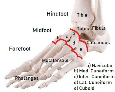
Learn about the 26 bones in each foot that are classified into tarsals, metatarsals, and phalanges. See labeled diagrams of the foot regions, joints, and muscles, ligaments, and tendons. Learn about the 26 bones and 33 joints in the foot, divided into hindfoot, midfoot and forefoot sections. See diagrams and pictures of the bones and their functions, such as weight-bearing, arch formation, and balance.

Anatomy Of The Foot Ankle Biology Diagrams
The foot is an intricate part of the body, consisting of 26 bones, 33 joints, 107 ligaments, and 19 muscles. Scientists group the foot's bones into into the phalanges, tarsal bones, and